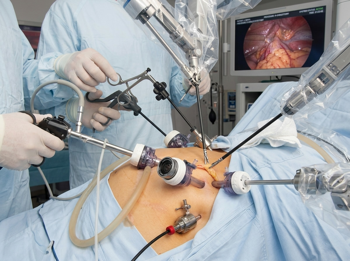
In modern HPB and general surgery, the Critical View of Safety (CVS) is widely accepted as the gold-standard method for preventing bile duct and vascular injuries during a laparoscopic cholecystectomy. First formalized by Strasberg and colleagues, achieving the CVS means confirming three concrete criteria before dividing any structures: clearing the hepatocystic triangle of all fibrofatty tissue, exposing the lower part of the cystic plate, and definitively demonstrating that two—and only two—structures enter the gallbladder.
When taught in textbooks or presented at conferences, the CVS is often depicted as a final destination—a neat, picturesque snapshot of anatomy where everything is isolated, skeletonized, and clear.
However, any experienced surgeon will tell you that focusing exclusively on the final picture misses the point. The CVS is a destination, but how you travel through the tissue to get there dictates the safety of the operation.
The Danger of Treating CVS as a “Checklist”
A common pitfall—particularly for surgical trainees—is treating the CVS as a visual checklist to be rushed through. When a surgeon views the CVS merely as an end-state, they can fall into the trap of aggressive dissection, forcing instruments into inflamed or unfamiliar tissue planes simply to make the final view “look right.”
In a hostile abdomen—such as a case of acute cholecystitis, severe fibrosis, or impacted stones—insistence on aggressively skeletonizing structures to achieve a textbook CVS can inadvertently cause the very injuries the concept was designed to prevent.
The CVS is not a method of dissection; it is a method of target identification. The safety of the procedure lies entirely in the careful, deliberate journey you take to uncover those structures.
Navigating the Journey: Safe Dissection Principles
Getting to the Critical View of Safety requires a disciplined approach to the anatomy. Safe travel through the hepatocystic triangle relies on several core principles:
- Respecting Tissue Planes: Dissection should be blunt and gentle where possible, using energy sources judiciously and keeping them well away from the main portal structures. Understanding natural fascial boundaries prevents thermal or mechanical trauma to the common bile duct.
- Working “On the Gallbladder”: A safe anatomical journey hugs the wall of the gallbladder. By staying close to the target organ—specifically dissecting the lower infundibulum off the cystic plate—you naturally distance your instruments from the hazardous common hepatic and bile ducts running deeper in the pedicle.
- Recognizing When to Change Route: A critical part of the journey is knowing when the path is blocked. If severe inflammation obscures the anatomy and the traditional path to the CVS cannot be safely mapped, a competent surgeon must pivot. This is where alternative routes or “bailout” strategies—such as a subtotal cholecystectomy or converting to open—become essential tools. Safety is defined by recognizing when the journey is too hazardous to continue as planned.
The Destination is Only as Good as the Path Taken
The Critical View of Safety provides intraoperative reassurance after it is established. But we must teach and practice it as an evolving process rather than a static snapshot.
The next time you are in the operating theatre performing a cholecystectomy, remember that the final view is only the reward. The real art of surgery happens in the quiet, methodical steps that get you there safely.